Using the latest in advanced imaging technology, a team of scientists from the United Kingdom, South Africa, and Spain performed a series of detailed examinations of the fossilized remains of Little Foot, a fossilized specimen from an extinct primate species that walked the earth 3.6 million years ago. Scrutinizing the most refined imagery available, the scientists behind the Little Foot study detected microscale-level details that revealed new data about Little Foot’s life and times, and about certain physical characteristics she possessed that may ultimately provide insights into the evolution of modern humans.
An Unprecedented Find
Little Foot was discovered by University of Witwatersrand paleoanthropologist Ron Clarke in 1994, during excavations in a cave near Johannesburg. Her remarkable state of preservation has allowed scientists to uncover many fascinating details about the physiology and lifestyle of her species, Australopithecus prometheus, which may have been an evolutionary forerunner of modern humans.

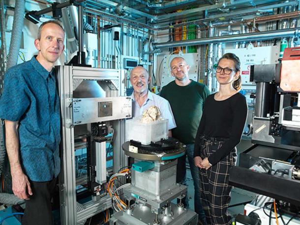
The Diamond Light Source team, which provided breakthrough scanning for the recently published Little Foot study. ( Diamond Light Source )
The Little Foot Study Relied On Synchrotron X-rays
To perform the crucial new tests for the Little Foot study the largely intact skeletal remains were secretly transferred out of South Africa in 2019, and brought to Britain’s national synchrotron X-ray facility, Diamond Light Source . Researchers at Diamond Light Source are pioneers in the use of intricately refined X-ray technology, which can help paleoanthropologists examine fossilized biological remains more deeply.
X-ray synchrotron scans can produce imagery that shows details down to a range of three micrometers. To put this in perspective, CT scans have long been considered state-of-the-art in medical scanning technology, but the resolution capacity of CT imagery is “only” 100 micrometers.
The study performed at Diamond Light Source focused on two areas of the Little Foot’s skeletal remains: the cranial vault, or upper section of the braincase, and the lower jaw, where the animal’s teeth were anchored. In each instance, significant revelations emerged.
Examinations of the cranial vault showed that tiny vascular canals, less than one millimeter wide, were still preserved and intact inside what would have been a soft section of skull bone when Little Foot was still alive.
According to University of Cambridge paleoanthropologist Amélie Beaudet, the chief architect of Diamond Light Source’s Little Foot study, these miniscule channels helped expedite the removal of excess heat from the Australopithecus prometheus brain, allowing for its release through the movements of the bloodstream.
The larger the brain, the more active and engaged the brain’s thermoregulation system must be, which is significant because the brains of modern humans are three times as big as those of Australopithecus prometheus.
If in fact Little Foot’s species was an evolutionary forerunner to humans, a closer study of its brain’s heat regulating system could reveal important facts about how this system would have changed over time to facilitate an evolutionary leap to Homo sapiens.
“This is very interesting as we did not have much information about that system,” Beaudet explained .
“Traditionally, none of these observations would have been possible without cutting the fossil into very thin slices,” she added. “But with the application of synchrotron technology there is an exciting new field of virtual histology [the study of microscopic structure of tissues].”

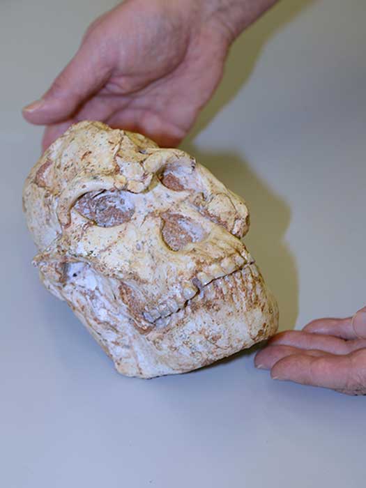
The Little Foot study was fortunate to have an excellent fossil specimen to work with. Shown here are her (Little Foot) teeth, which look pretty good even 3 million years later! ( Diamond Light Source )
The Study Also Looked At Her Fossilized Teeth
Examination of Little Foot’s teeth didn’t reveal much about the female creature’s evolutionary path. But it did show that her life was far from easy.
“In the teeth, we can see some defects, like lines or grooves,” said Beaudet . “It means at some point the enamel could not form properly.” This disruption in enamel formation would most likely have been caused by something traumatic she experienced during childhood, when her teeth were still in the process of growing and developing.
It is possible that Little Foot was deprived of food as a child, which resulted in malnutrition. This may have been caused by environmental or climatological changes that created food scarcity among her social group.
“We know that the environment was not always stable,” Beaudet said.
Another possibility is that Little Foot suffered from some type of serious illness when she was young, of a type that would interfere with her normal growth.
Both of these explanations contain a degree of speculation, which is impossible to avoid when trying to interpret the life experiences of a primate that lived three million years ago.
“We cannot say [if] it was because of a food shortage, or because she was sick, or something else,” Beaudet declared. There is no doubt Little Foot experienced hardship, but the true nature of her struggles will remain a puzzle that eludes a definitive conclusion.
Little Foot’s foot bones. (Photograph by Mike Peel ( www.mikepeel.net) / CC BY-SA 4.0 )
Solving Evolutionary Mysteries With Fossils And High Tech
Little Foot was much smaller than a modern human. She was just 51 inches (130 centimeters) tall, despite being a full-grown adult. Her features included a mixture of characteristics common to humans and other primates, and the shape of her skull and face were decidedly ape-like.
While Little Foot’s species might have been an evolutionary forerunner to humans, it also might have represented an evolutionary dead end, similar to Neanderthals. Modern scanning technology that allows experts to examine fossil evidence down to the minutest detail could help them make such a distinction, with respect to Australopithecus prometheus and any other extinct species of primate that produces a fossil.
“Important aspects of early hominin biology remain debated, or simply unknown,” noted Diamond Life Source scientist Dr. Thomas Connolley. “In that context, synchrotron X-ray imaging techniques like microtomography have the potential to non-destructively reveal crucial details on the development, physiology, biomechanics and taxonomy of fossil specimens.”
Little Foot’s nearly intact fossilized remains offered scientists a perfect opportunity to test the capacities of this type of technology.
“This level of resolution is providing us with remarkably clear evidence of this individual’s life,” marveled principal investigator Dominic Stratford from the University of Witwatersrand, who accompanied Little Foot to the United Kingdom from South Africa. “We think there will also be a hugely significant evolutionary aspect, as studying this fossil in this much detail will help us understand which species she evolved from and how she differs from others found at a similar time in Africa.”
Synchrotron X-ray scanning, which was really the breakthrough in the Little Foot study, is opening exciting new vistas for paleoanthropologists and others who study fossilized biological remains.
Significant scientific discoveries that resolve many unanswered questions may result from a wider application of this breakthrough technology.
Top image: The biggest breakthrough in the Little Foot study was the scientific power of Diamond Light Source’s advanced synchrotron technology. Source: Diamond Light Source
By Nathan Falde
Related posts:
Views: 0
 RSS Feed


 March 4th, 2021
  Awake Goy
  Posted in
  Tags:
Tags: 


